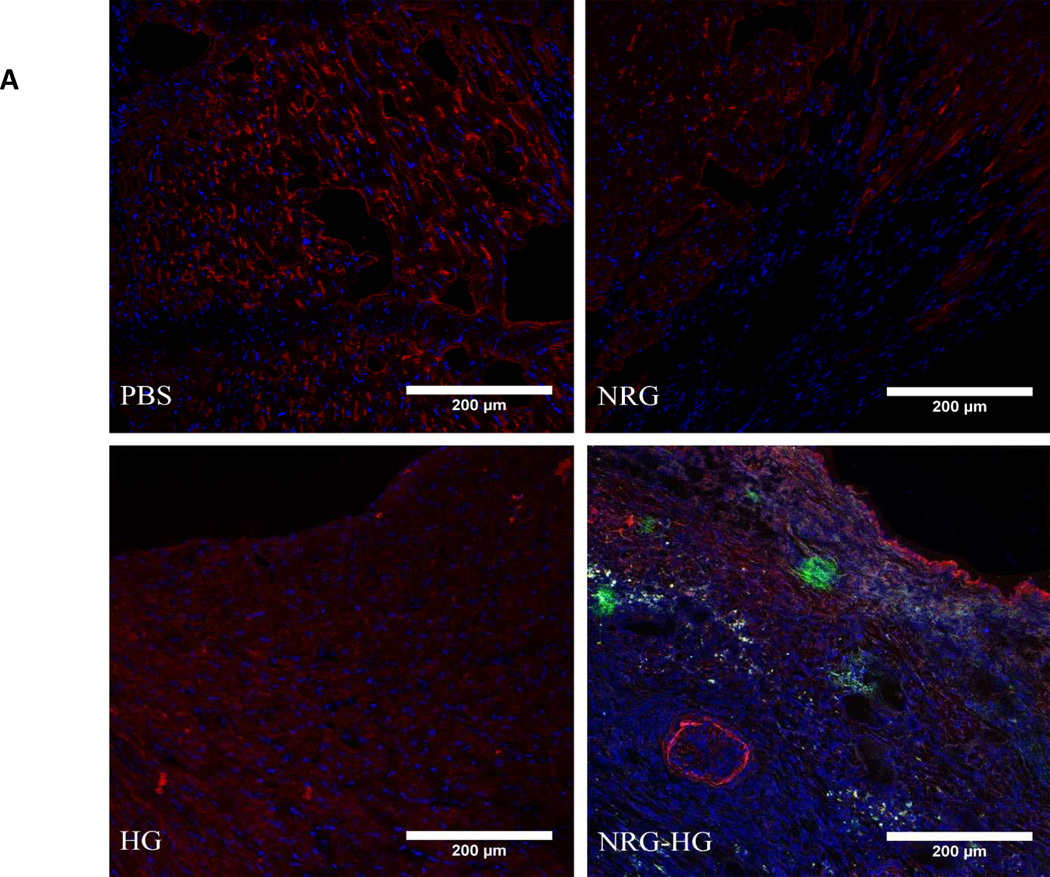
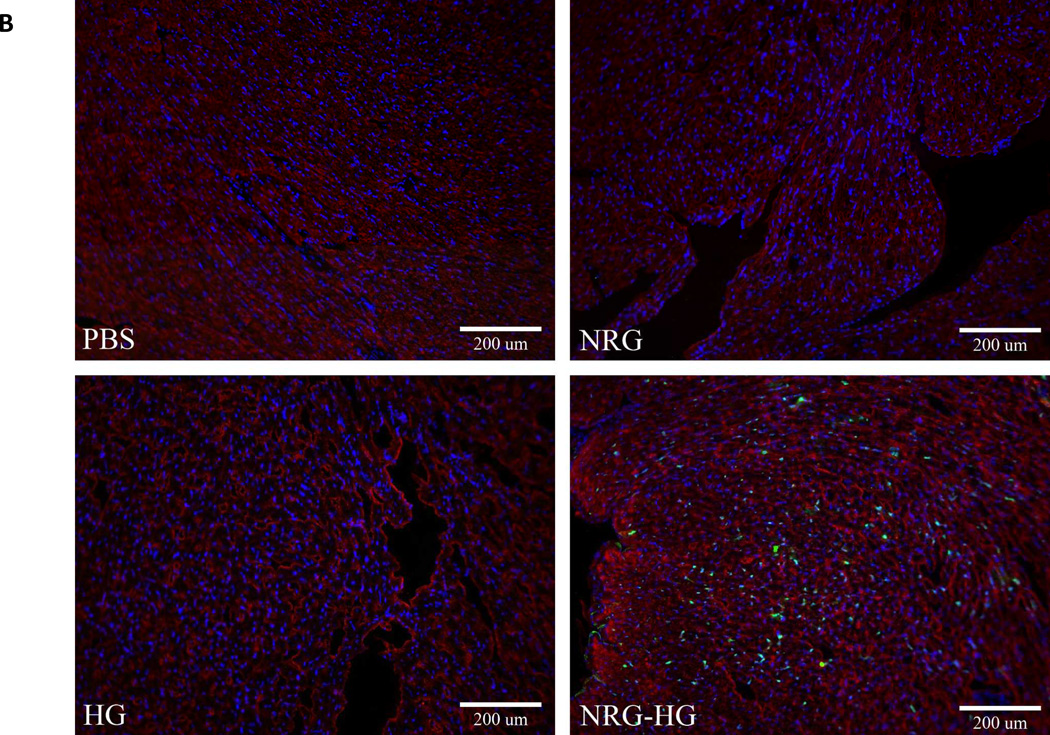
Figure 3. Mitotic activity in peri-infarct region.
Heart sections at 6 days following treatment visualized under fluorescent microscopy demonstrating PH3 and Ki67 positivity with NRG-HG treatment. Sections are stained with DAPI (blue), troponin (red), PH3 (A) and Ki67 (B) (green). All animals treated with NRG-HG demonstrated presence of PH3 and Ki67 in the peri-infarct region.


