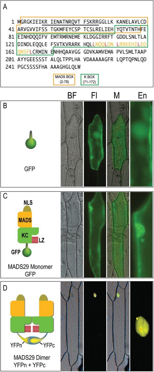
Fig. 1.

Intracellular localization of M29 monomer and homodimer. (A) M29 polypeptide sequence showing the position of conserved domains and motifs. The MADS, and K-box domains are marked by different coloured boxes. The unmarked region represents the C-terminal domain. The NLS sequence is underlined and the leucine-zipper-like motif in the K-box has been shown by yellow font with five conserved leucine residues marked in red. (B–D) The left subpanels in panels B, C, and D contain graphical representations of the M29:reporter constructs used in this experiment, while the actual expression of these chimeric proteins in onion epidermal cells is shown in the respective right subpanels. (B) GFP (C) M29:GFP (D) M29:YFPc+M29:YFPc. BF, bright field; Fl, fluorescence image; M, Merged image; En, enlarged view of the nuclear region.
