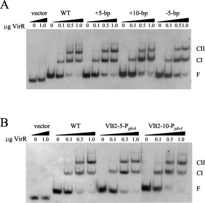
FIG. 2.
Gel mobility shift analysis of altered VirR box regions. Each 183-bp DIG-labeled DNA target was incubated in the presence of various amounts of purified VirR, as indicated above each lane. Note that 1 μg of VirR represents a final concentration of 1.55 μM. The free DNA (F), CI, and CII bands are labeled. The wedges above the lanes are used to distinguish the separate DNA targets. (A) WT, +5-bp, +10-bp, and −5-bp indicate wild-type VirR boxes and VirR boxes containing a 5- or 10-bp insertion or a 5-bp deletion, respectively. (B) WT, VB2-5-PpfoA, and VB2-10-PpfoA indicate wild-type VirR boxes and DNA targets containing a 5- or 10-bp insertion between VirR box 2 and the promoter, respectively.

