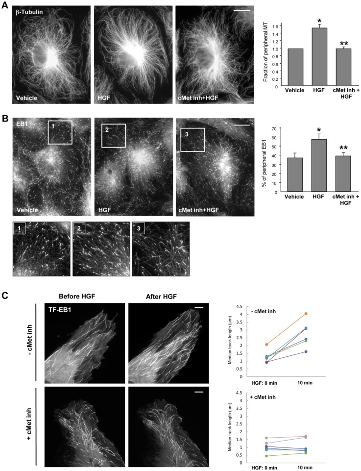
Figure 1. HGF stimulates peripheral MT growth.
HPAEC grown on coverslips were stimulated with HGF (50 ng/ml, 10 min) with or without pretreatment with c-Met inhibitor (carboxamide 50 nM, 30 min) followed by A: Immunofluorescence staining with an antibody against β-tubulin; B: Immunostaining with anti-EB1 antibody. Insets show high magnification images of cell periphery areas with microtubules or EB1-positive microtubule tips. Bar = 5 µm. Results are representative of five independent experiments. Bar graphs depict results of quantitative analysis of peripheral microtubules (A, right panel) and peripheral EB1 (B, right panel) in methanol-fixed HPAEC; *P<0.05; n = 4; 6 images from each experiment. C: Live cell imaging of HPAEC expressing GFP-EB1 stimulated with HGF with or without pretreatment with c-Met inhibitor. Projection analysis of 20 consecutive images before and after HGF treatment shows changes in GFP-EB1 track length. Bar = 2 µm. Quantification of GFP-EB1 track length is presented on right panels. Each pair of dots represents the median track length in a cell before and after thrombin treatment. Results are representative of four independent experiments; eight cells have been inspected for each condition, in each experiment.

