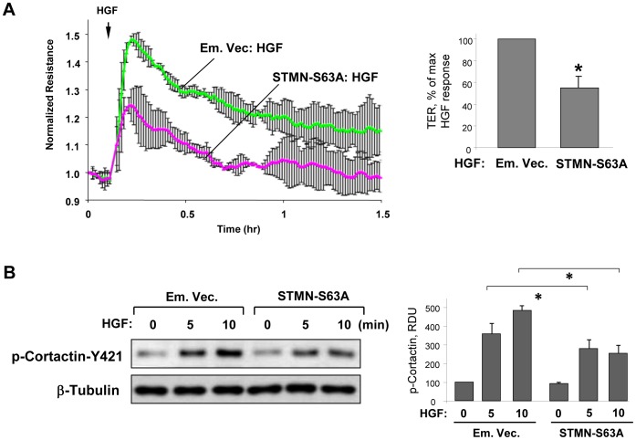
Figure 4. Expression of phosphorylation-deficient stathmin attenuates HGF-induced EC barrier enhancement.
A: Endothelial monolayers transfected with phosphorylation-deficient stathmin (STMN-S63A) or empty vector (Em. Vec.) were stimulated with HGF (50 ng/ml). A: TER measurements were performed over 1.5 hrs. Bar graphs depict results of quantitative analysis of permeability data; n = 5; *P<0.05. B: Cortactin phosphorylation at Y421 and tubulin acetylation at indicated time points of HGF treatment was monitored by Western blot. Probing for β-tubulin was used as a normalization control. Results are representative of three independent experiments. Bar graphs depict the quantitative densitometry analysis of western blot data; n = 4; *P <0.05, RDU: relative density units.

