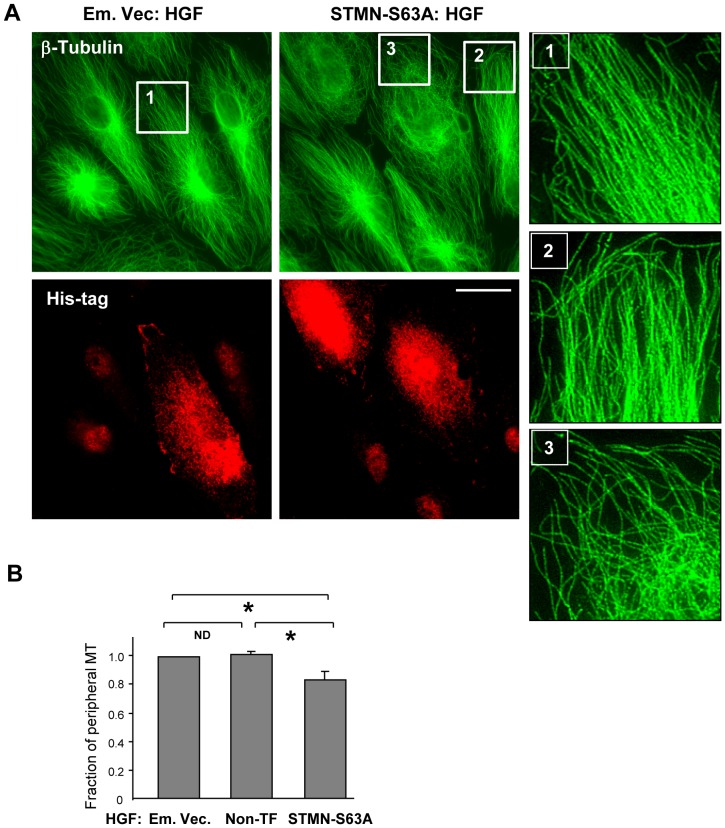
Figure 5. Expression of phosphorylation-deficient stathmin attenuates HGF-induced stimulation of peripheral MT network formation.
Cells grown on coverslips were transfected with empty vector (Em. Vec.) or STMN-S63A and stimulated with HGF (50 ng/ml, 10 min). A: MT network was visualized by immunofluorescence staining of methanol-fixed cells with an antibody against β-tubulin. Transfected cells were detected by staining with His-tag antibody. Insets show magnified images with details of MT structure in non-transfected and STMN-S63A transfected cells. Bar = 10 µm. Results are representative of three independent experiments. B: Bar graphs depict results of quantitative analysis of peripheral microtubules; n = 3; 10 cells from each experiment; *P<0.05.

