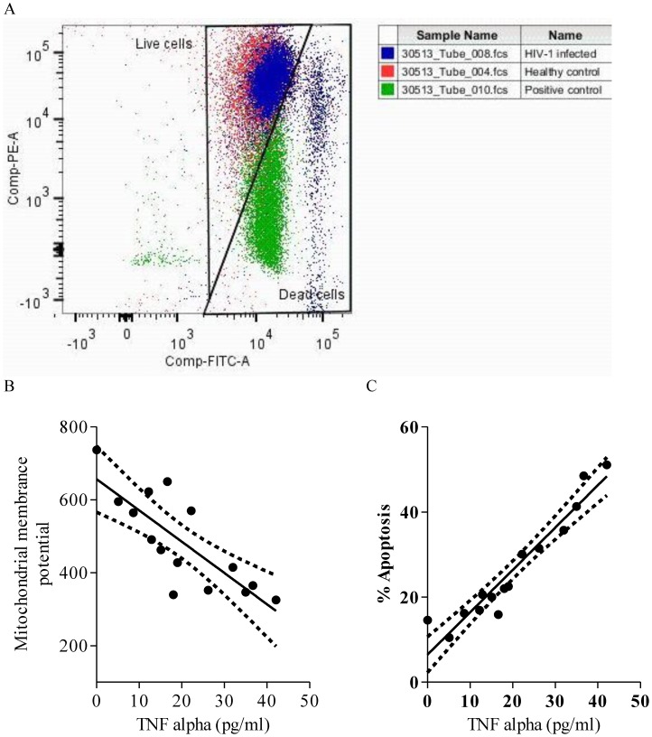
Figure 8. Higher plasma TNF-α levels up-regulate apoptosis of peripheral lymphocytes in HIV-1 infected individuals.
A. Representative overlay dot plot showing JC-1 staining of peripheral blood lymphocytes depicting a shift of fluorescence emission from red (FL2) to green (FL1) indicating loss of mitochondrial membrane potential, which is associated with apoptosis in HIV-1 infected individuals (blue). Negative control (red, healthy control) and positive control (green, protonophore FCCP treated) were used to set the gate for dead cells. B. Representative scatter plots depicting significant negative correlation between TNF-α expression and mitochondrial membrane potential (ΔΨm) in HIV-1 infected individuals. C. Representative scatter plots depicting significant positive correlation between TNF-α expression and cells undergoing apoptosis in HIV-1 infected individuals. Solid line represents linear regression of points and dotted lines represents 95% confidence band for their mean values. Rho and p-values are from the Spearman rank correlation test.

