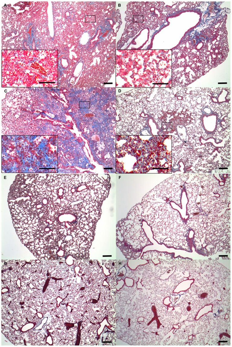
Figure 3. Histopathological findings of fibrosis induced by bleomycin.
Representative results of Masson's trichrome staining of lung tissue from mice in the bleomycin-induced lung injury model (A, B, C and D). Masson's trichrome staining of lung tissue from (A) wild-type (WT) and (B) knockout (KO) mice on the seventh day after intratracheal administration of bleomycin. Masson's trichrome staining of lung tissue from (C) WT and (D) KO mice on the 28th day after intratracheal administration of bleomycin. All scale bars = 100 µm. The window shows an area of increased magnification revealing a part of lung fibrosis. Representative results of Masson's trichrome staining of lung tissue from mice in the saline-treated control group (E, F, G and H). Masson's trichrome staining of lung tissue from (E) WT and (F) KO mice on the seventh day after intratracheal administration of saline. Masson's trichrome staining of lung tissue from (G) WT and (H) KO mice on the 28th day after intratracheal administration of saline.

