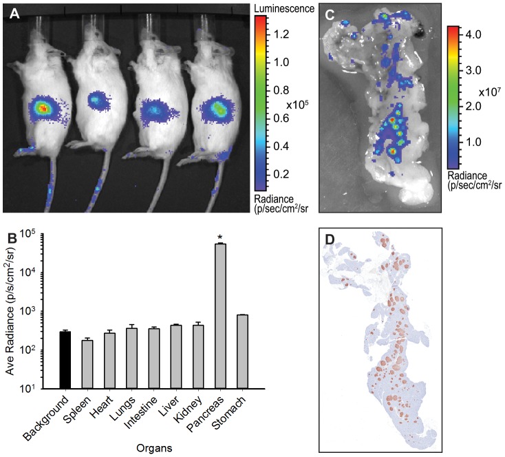
Figure 1. Luciferase expression is restricted to the pancreas.
A) An example of BLI on ob/ob-luc mice. The animals of the same age were injected i.p. with D-luciferin and imaged 5 min later. Signal is present in the midsection of the animals only. B) Biodistribution of luciferase in the organs of ob/ob-luc mice. Three animals with similar intensities in the midsection were dissected and the organs were imaged with an IVIS Spectrum. ROI analysis was performed on the organs. The background bar is highlighted to show all organs except pancreas are at background levels while pancreas is significantly higher than other organs (*p<0.001, One way ANOVA). C) Zoomed in BLI of a pancreas. D) The pancreas was sectioned and stained with anti-insulin antibody. Both BLI and insulin staining have similar punctate patterns.

