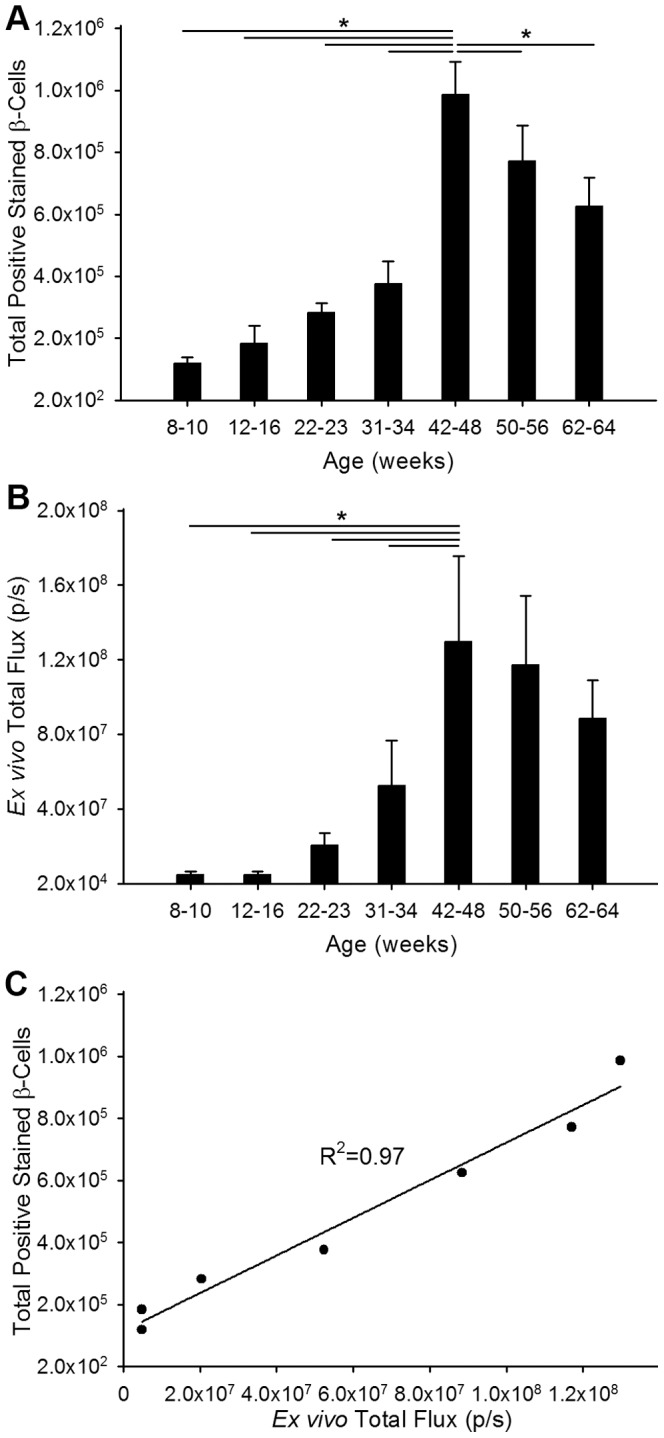
Figure 4. Correlation of ex vivo bioluminescence with quantitative histology.

A) Total number of β-cells was counted from sections of pancreas from ob/ob-luc mice (N = 3–8/group). Mice were grouped to minimize variation since individual mice reach disease stages at different time points. B) Total bioluminescence was measured from ex vivo images of pancreata from ob/ob-luc mice. When the data are grouped similar to β-cell number there is an upwards trend in β-cell number as the mice age. (The horizontal lines in figures A and B indicate a statistically significant difference between the two groups, *p<0.001, One way ANOVA) C) There is a strong correlation between the number of β-cells counted by histology and ex vivo BLI measurements.
