Figure 1.
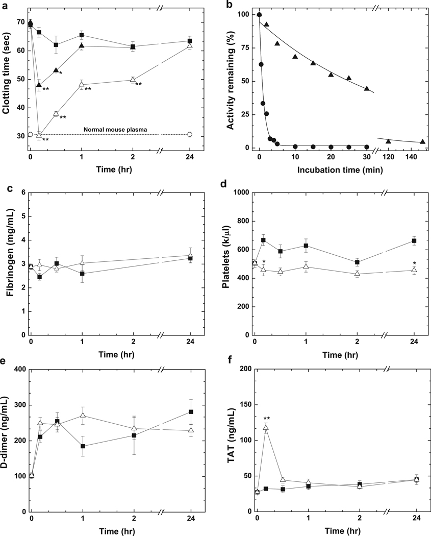
Effect of hFXaI16L on coagulation parameters. In a, HB mice (Balb/c) were injected with PBS (-■-; n = 7) or hFXaI16L (-▲-, 225 µg/kg, n = 4; -△-, 450 µg/kg, n = 9). At the indicated time intervals a modified one-stage aPTT was performed on processed mouse plasma. Clotting times for wt-mice (-○-, n = 7) is indicated. In b, wt-hFXa (-●-, 20 nM) or hFXaI16L (-▲-, 20 nM) were incubated in diluted HB (Balb/c) mouse plasma and residual activity was assessed by clotting assay. The solid lines were drawn following analysis of data sets to a single exponential decay with fitted half-lives of: wt-hFXa, 0.15 ± 0.01 min; hFXaI16L, 5.3 ± 0.46 min. These values have been appropriately adjusted for sample dilution. The data are representative of two to three similar experiments. In c–f, markers of coagulation activation were measured. HB mice (Balb/c) were injected with PBS (-■-; n = 3–10) or hFXaI16L (-△-, 450 µg/kg, n = 3–8). At the indicated time intervals, levels of c) fibrinogen, d) platelets, e); D-dimer and f) TAT complex were measured. In panels a and c–f, all measurements are presented as mean ± SEM. For statistical comparisons, treated animals are compared to HB-PBS controls at the same time point; ** p<0.001; * p<0.05.

