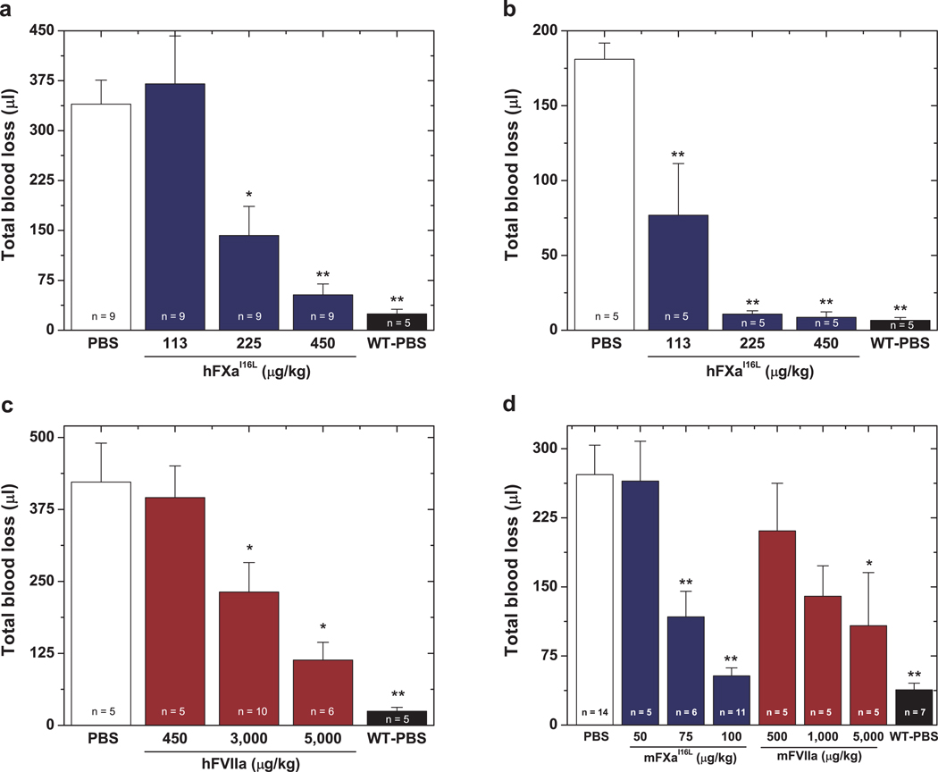
Figure 2.
Blood loss following tail-clipping. In a, five minutes prior to injury, PBS (white column) or hFXaI16L (blue columns) was administered to HB mice (C57Bl/6) via tail vein at the indicated dosage. Total blood loss (µl) was then measured following tail transection. In b, tail clipping was performed on wt-mice or HB mice (Balb/c) prior to protein infusion and blood was collected for 2 min (total blood loss at 2 min: wt-mice: 17 ± 5.4 µl; HB: 49 ± 7.7 µl). Subsequently, PBS (white column) or hFXaI16L (blue columns) was administered to HB mice via a pre-inserted jugular vein cannulus at the indicated dosage; wt-mice also received PBS (black column). Blood was collected for an additional 10 min in a fresh tube of saline and total blood loss (µl) was measured. Data in (b) represent total blood loss subsequent to protein infusion. In c, five minutes prior to injury, PBS (white column) or hFVIIa (red columns) was administered to HB mice (C57Bl/6) via tail vein at the indicated dosage. Total blood loss (µl) was then measured following tail transection. In d, five minutes prior to injury, PBS (white column), mFXaI16L (blue columns), or mFVIIa (red columns) was administered to HB mice (Balb/c) via tail vein at the indicated dosage. Total blood loss (µl) was then measured following tail transection. In a–d, hemostatically normal mice on the appropriate strain infused with PBS (WT-PBS, black column) served as a control. In each panel, the number of animals per group is indicated and all measurements are presented as mean ± SEM. For statistical comparisons, treated animals are compared to HB-PBS controls ** p<0.001; * p<0.05.

