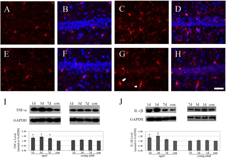
Figure 4. Hepatectomy differentially induced strong neuroinflammation at hippocampus of aged and adult rats.
A: Iba1 staining (red) of CA1 at normal young adult rat. B: the merged panel of Iba1 staining (red) and Dapi (blue) of CA1 at normal young adult rat. C: Iba1 staining (red) of CA1 at young adult rat 1d after surgery. D: the merged panel of Iba1 staining (red) and Dapi (blue) of CA1 at young adult rat 1d after surgery. E: Iba1 staining (red) of CA1 at normal aged rats. F: the merged panel of Iba1 staining (red) and Dapi (blue) of CA1 at normal aged rats. G: Iba1 staining (red) of CA1 at aged rat 1d after surgery. Activated microglia (white arrow) was observed. H: the merged panel of Iba1 staining (red) and Dapi (blue) of CA1 at aged rat 1d after surgery. I: western blot of TNF-α at hippocampus. J: western blot of IL-1β at hippocampus. Data were mean ± SD. *p<0.05 vs control. Bar = 50 um.

