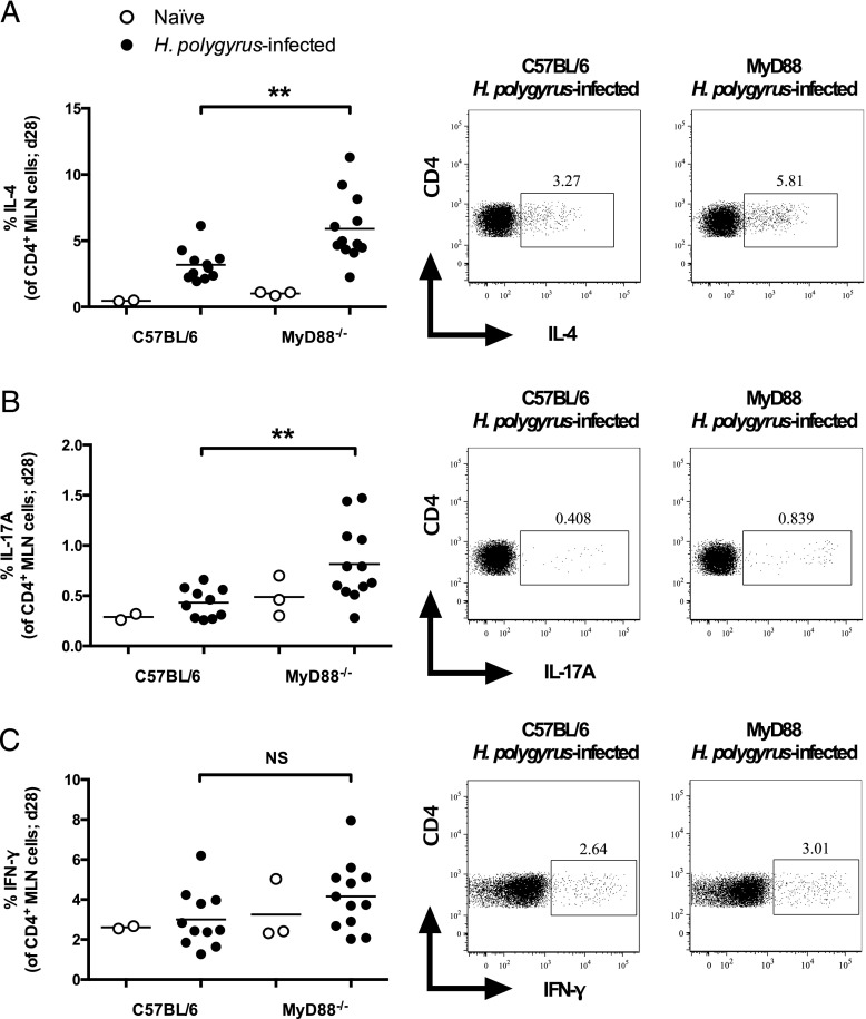
FIGURE 3.
MyD88−/− mice mount stronger CD4+ T cell IL-4 and IL-17A responses following H. polygyrus infection. C57BL/6 and MyD88−/− mice were left naive or infected with 200 H. polygyrus third stage larvae. At 28 d following infection, MLN cells were isolated and restimulated with PMA/ionomycin and brefeldin A, after which cells were stained as indicated and cytokine production was measured by flow cytometry. (A) Percentage of IL-4–producing cells among CD4+, live lymphocyte cells, and representative dot plots. (B) Percentage of IL-17A–producing cells among CD4+, live lymphocyte cells, and representative dot plots. (C) Percentage of IFN-γ–producing cells among CD4+, live lymphocyte cells, and representative dot plots. Data shown in (A)–(C) are pooled from two independent experiments, each with four to seven H. polygyrus-infected mice per group; naive mice were examined in only one of these experiments. Statistics shown indicate comparisons between infected groups. **p ≤ 0.01.

