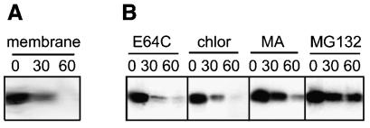
FIG. 4.
Degradation of p10 occurs following membrane insertion and is altered by a proteasome inhibitor. (A) Pulse-chase analysis for the indicated times (in minutes) was performed with p10-transfected cells as described in the legend to Fig. 1. After the pulse or each chase, the membrane fraction was isolated, and the presence of membrane-associated p10 was determined by immunoprecipitation, SDS-PAGE, and fluorography. (B) Pulse-chase analysis for the indicated times (in minutes) was performed with p10-transfected cells as described in the legend to Fig. 1, except that cells were incubated during the pulse-chase in the presence of inhibitors of lysosomal proteases (E64C) or lysosome acidification (chloroquine [chlor] or methylamine [MA]) or a proteasome inhibitor (MG132).

