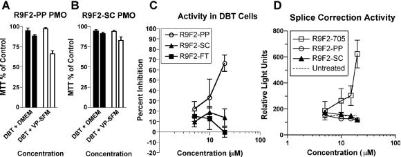
FIG. 2.
Cytotoxicity of the R9F2-PP PMO (A) and R9F2-SC PMO (B) was tested by MTT assay at 10 μM (left bar of each pair) or 20 μM (right bar of pair) concentration. The means of three experiments with standard errors are shown. Cells transfected with a plasmid containing a MHV target gene-luciferase fusion were assayed for inhibition of luciferase expression after treatment with R9F2-PMO (C). DBT cells transfected with a plasmid containing a missplicing luciferase reporter gene were assayed in a splice correction assay after treatment in cell culture medium containing R9F2-PMO (D). Relative light units produced from translated luciferase in each monolayer are shown. The dotted line represents the mean of nine controls treated with medium alone. Error bars (C and D) represent the standard errors of the means of three replicates.

