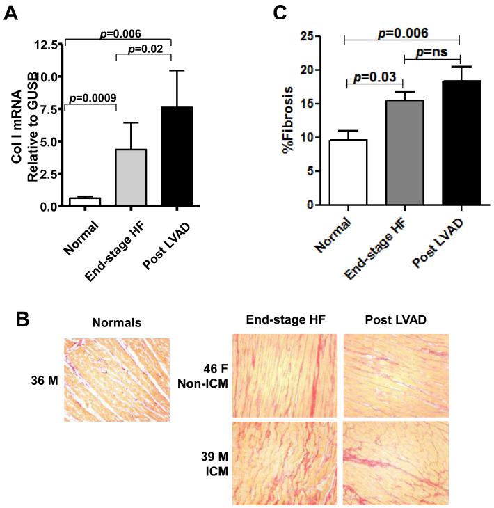
Figure 1. Fibrosis in normal, end-stage HF and post LVAD LV tissues.
A:Collagen (Col) I mRNA expression in normal, end-stage HF, and post LVAD LV tissues. B: Representative Picrosirius Red staining at × 20 objective magnification. 36 M, 36 years old male; Non-ICM, non-ischemic cardiomyopathy; 46 F, 46 years old female; 39 M, 39 years old male. C: Quantification of Picrosirius Red staining for interstitial Col deposition for normal, end-stage HF and post LVAD LV tissues.

