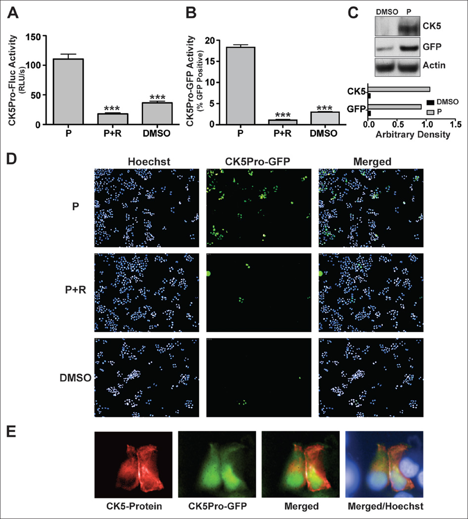
Figure 1.
CK5 expression in response to progesterone in T47D cells. CK5 promoter activity was assessed by (A) luciferase reporter assay of CK5Pro-Fluc-T47D cells and (B) green fluorescent protein (GFP) reporter assay of CK5Pro-GFP-T47D cells in the presence of 100 nM progesterone (P) or progesterone plus 1 µM RU486 (R) and DMSO (vehicle for both P and R). (C) CK5Pro-GFP-T47D Western blot analysis of CK5 and GFP protein expression from cells treated with 100 nM progesterone. Arbitrary density was obtained by Image J software. (D) Representative images of CK5Pro-GFP-T47D cells treated with 100 nM progesterone or 100 µM progesterone plus 1 µM RU486. (E) CK5 and GFP protein expression as assessed by immunofluorescence assay. CK5Pro-Fluc-T47D cells treated with 100 nM progesterone were stained with anti-CK5 antibody (red), and GFP-expressing cells are green. Nuclei were stained with Hoechest 33342 (blue). Student’s t-test was used for statistical analysis with ***p ≤ 0.001.

