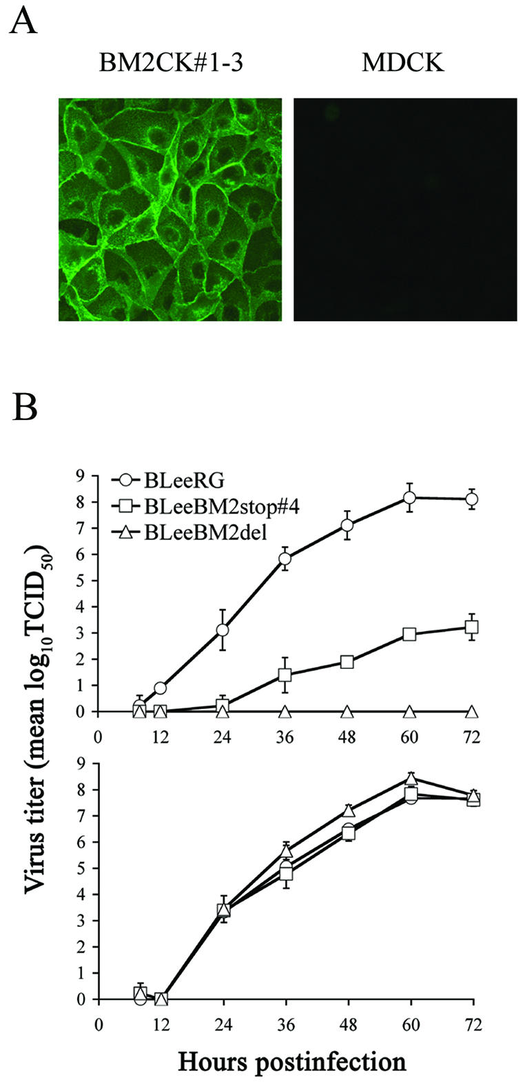
FIG. 6.

Generation of cells constitutively expressing the BM2 protein and growth curves for BLeeRG and mutant viruses in MDCK cells and BM2-expressing cells. (A) BM2CK#1-3 (left panel) and MDCK cells (right panel) were fixed with 3% formaldehyde solution and permeated with 0.1% Triton X-100. The BM2 protein was detected with rabbit anti-BM2 peptide serum, used as a primary antibody, and FITC-conjugated anti-rabbit IgG as a secondary antibody. (B) MDCK and BM2CK#1-3 cells were infected with virus (103 TCID50) which had been amplified in BM2CK#1-3 cells. At the indicated times after infection, the titers of virus in the supernatant from infected MDCK (upper panel) and BM2CK#1-3 (lower panel) cells were determined with BM2CK#1-3 cells. The values are means (± standard deviation) of three determinations.
