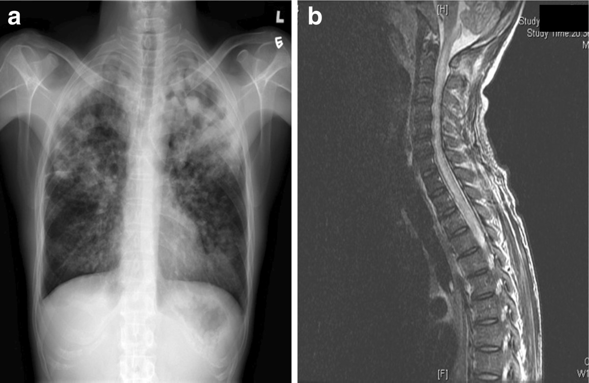
Figure 1.

Radiological imaging of lung and spinal cord. a: Chest X-ray (PA Erect film) on initial presentation showing bilateral lung field patchy consolidation with left upper zone predominance. b: Sagittal section of MRI spine showing diffuse T2-weighted hyperintensity involving C1-T4 levels of the spinal cord suggestive of longitudinally extensive transverse myelitis (LETM).
