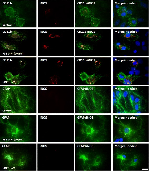
Figure 7.

Cellular localization of inducible nitric oxide synthase (iNOS) in lipopolysaccharide (LPS) cultures. Cells were incubated with UDP or PSB 0474 for 48 h. Microglia were labeled with mouse anti-CD11b (Alexa Fluor 488, green), astrocytes with mouse anti-GFAP (Alexa Fluor 488, green) and iNOS with rabbit anti-iNOS (TRITC, red). Cell nuclei were labeled with Hoechst 33258 (blue). The orange spots represent the expression of iNOS in the cells and are coincident with an increased expression of iNOS in microglia, but not in astrocytes, upon stimulation with the uracil nucleotides. Scale bar = 10 μm.
