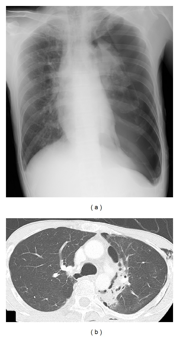
Figure 1.

Chest X-ray performed at initial presentation and chest computed tomography on the 11th hospital day. (a) On admission, chest X-ray showed a left-sided pneumothorax. (b) On the 11th hospital day, chest computed tomography showed intrapulmonary infiltration on the mediastinal side of the left upper lobe, and no emphysematous changes were seen.
