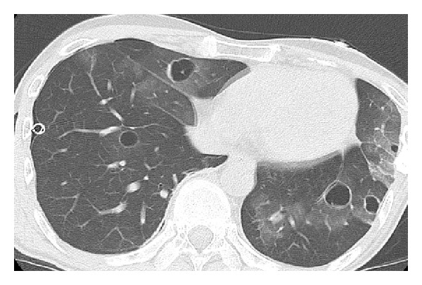
Figure 3.

Chest computed tomography taken 9 months after the operation for pulmonary complications shows bilateral thin-walled cystic lesions that are surrounded by ground glass opacities, for which the patient underwent placement of a drainage tube in the pleural space for right pneumothorax.
