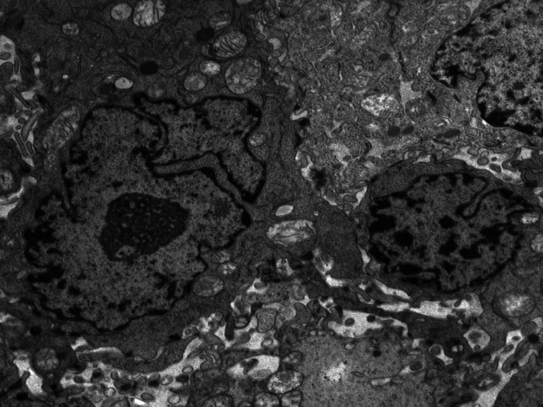
Fig. 5.

The micrograph presents differences between the nuclei of tumor cells. One is much larger, with a deeply invaginated nuclear membrane, enlarged active nucleolus, a field of interchromatin granules, and small groups of heterochromatin located in the vicinity of the nuclear membrane
