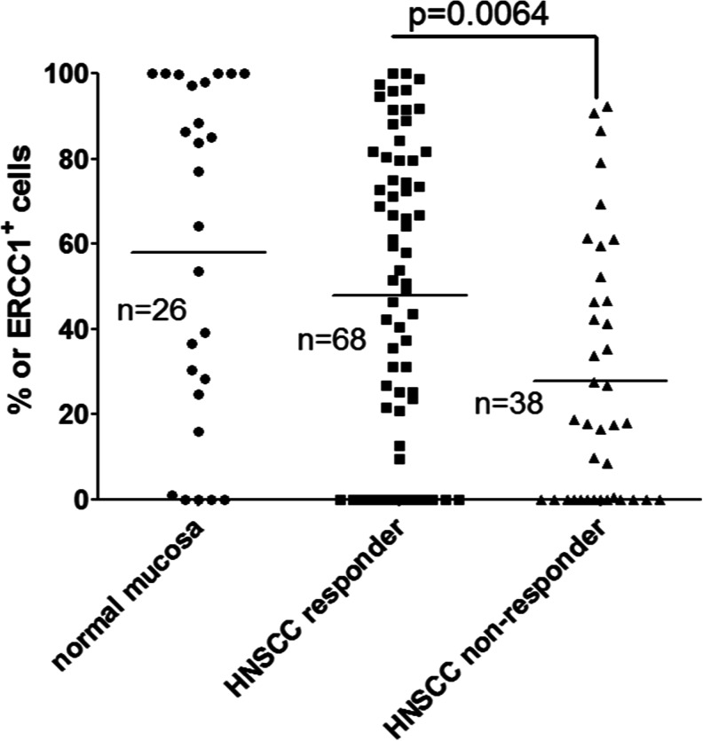
Fig. 1.
Twenty-six normal mucosa samples and 106 HNSCC tumor tissue samples have been stained with 8 F1 ERCC1 antibody, and the percentage of stained cells in tumor cell nests or in high power fields of normal mucosa has been evaluated. There was no significant difference between the staining representation of normal mucosa and therapy responder HNSCC, but the therapy nonresponder HNSCC patients have shown significantly lower (p = 0.0064) ERCC1 representation compared to the normal mucosa and responder patients

