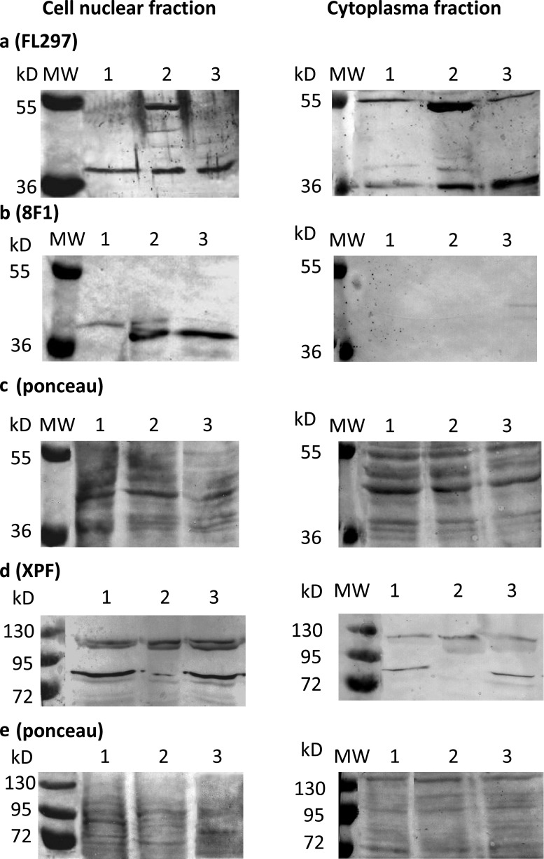
Fig. 3.
Western blot of FL-297 (a) and 8 F1 (b) anti-ERCC1 antibodies and of SPM228 (d) anti-XPF antibody in SCC-25 (lane 1) and Detroit 562 (lane 2) squamous cell carcinoma cell lines and in not transformed BEAS-2B bronchial epithelial cells (lane 3) using cell nuclear (left panels) and cytoplasm (right panels) fractions. Ponceau-stained whole protein detection was used as loading control (c, e)

