Fig. 5.
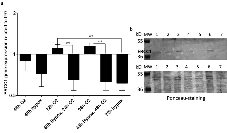
Detroit 562 cells were cultured in hypoxic and normoxic conditions for 48–96 h and ERCC1 mRNA expression was examined (a), and western blot of nuclear ERCC1 protein (b) was performed. The ERCC1 mRNA expression (a) was normalized with β-actin gene expression and was proportioned to the gene expression of control zero (the gene expression of the t = 0 days, which was the reference for time and treatment-related changes). “1” represents no change compared to start time (without treatment). The normoxic culture showed no significant fluctuation of ERCC1 gene expression, while the hypoxic culture (even if followed by normoxic) resulted in significant ERCC1 gene expression decrease. Western blot of 8 F1 (b, upper panel) anti-ERCC1 antibody in cell nuclear extracts of Detroit 562 squamous cell carcinoma cell line cultured 48 h normoxic (1), 48 h hypoxic (2), 72 h normoxic (3), 72 h hypoxic (4), 48 h hypoxic and 24 h normoxic (5), 96 h normoxic (6), and 48 h hypoxic and 48 h normoxic (7). Ponceau-stained whole protein detection in nuclear extracts was used as loading control (b, lower panel). Hypoxic culture conditions, even when followed by normoxic periods, lead to significant reduction of ERCC1 gene expression at mRNA and protein levels

