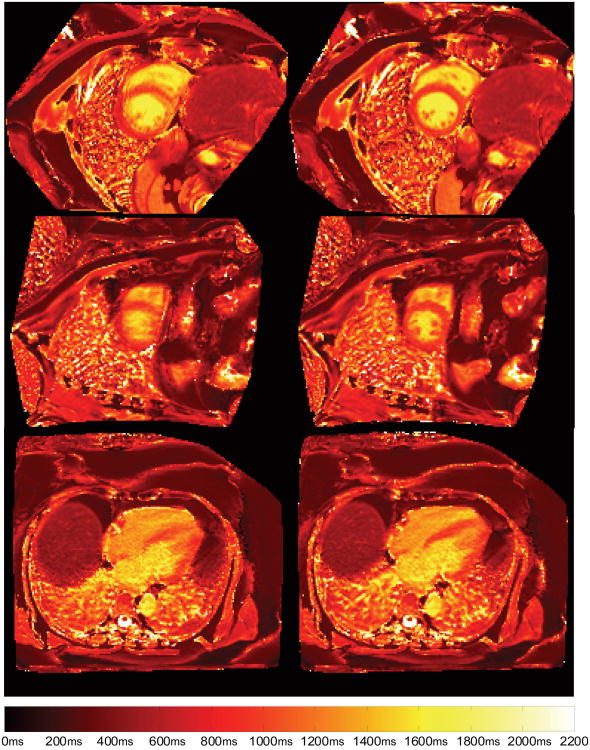
FIG 10.
Example of improved T1 mapping after motion correction. Left column: T1 maps of original MOLLI images indicating smearing on the myocardium due to imperfect breath-holding. Right column: Sharp myocardial boundary is recovered after motion correction using proposed technique. These images were from 3 different patients.

