Figure 5:
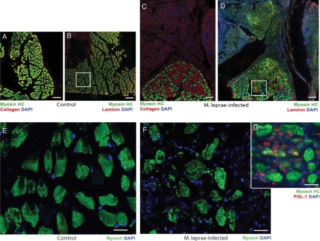
Pathological features of skeletal muscles in infected armadillos. (A-D) Double immunolabeling of transverse sections of control (A, B) and infected (C, D) deep lumbrical muscles showing an accumulation of fibrotic tissues (C, D) and immunolabeling with antibodies to myosin heavy chain (Myosin HC; green) and laminin or collagen (red), counter stained with DAPI for nuclei (blue). Note the massive number of nucleated cells present in fibrotic tissues associated with infected muscles that were not present in the control muscles (weak myosin HC staining in green in fibrotic tissues represent non-specific background fluorescence). Also note the disorganized muscle architecture and reduced number of muscle fibers with accumulated matrix in infected muscles (C and D) as compared to controls (A and B). Higher magnification of myosin HC in the boxed areas in (B) and (D) are shown in (E), (control: myosin HC in green), (F) (infected: myosin HC in green) and (G) (infected: myosin HC in green and M. leprae labeling by PGL-1 antibody in red counter stained with DAPI for blue nuclei). Note the disorganized myosin HC labeling showing disrupted muscle fibers with infiltrated cells (blue nuclei) containing large number of M. leprae (red labeling in G) infected animals.

