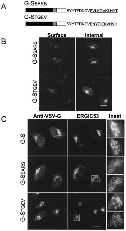
FIG. 6.
Group 1 coronaviruses and SARS S proteins contain intracellular localization signals in their cytoplasmic tails. (A) The final 11 amino acids of TGEV S or SARS S (underlined), which contain putative dibasic signals, were swapped for the same residues of G-S. (B) Intact HeLa cells expressing G-SSARS or G-STGEV were stained with mouse anti-VSV-G at 4°C, fixed, permeabilized, and then stained with rabbit anti-VSV-G for internal expression. Secondary antibodies were fluorescein-conjugated donkey anti-rabbit IgG and Texas Red-conjugated goat anti-mouse IgG. (C) HeLa cells expressing G-S, G-SSARS, or G-STGEV were fixed, permeabilized, and stained with rabbit anti-VSV-G and mouse anti-ERGIC-53. Secondary antibodies were fluorescein-conjugated goat anti-rabbit IgG and Texas Red-conjugated donkey anti-rabbit IgG. The insets show enlargements of the boxed regions, with the anti-VSV-G panel on top and the anti-ERGIC-53 panel below. Bars, 10 μm.

