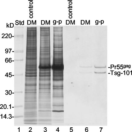
FIG. 8.
Concentration of Tsg-101 into Gag particles. Donor membranes (DM) and Gag particles (g-p) from BHK-21 cells infected with SFV-C/Pr55gag vectors and pre- and postlabeled with [35S]Met as well as donor membranes from uninfected control cells were analyzed by SDS-PAGE on an equal-lipid basis. The proteins in the gel were transferred to a filter, and this was used for the detection of Tsg-101 by Western blotting (lanes 5 to 7). Lanes 2 to 4 represent an autoradiograph of 35S-labeled proteins separated on a duplicate gel. Note that abundant Pr55gag was stained nonspecifically in the blot. Std, standard.

