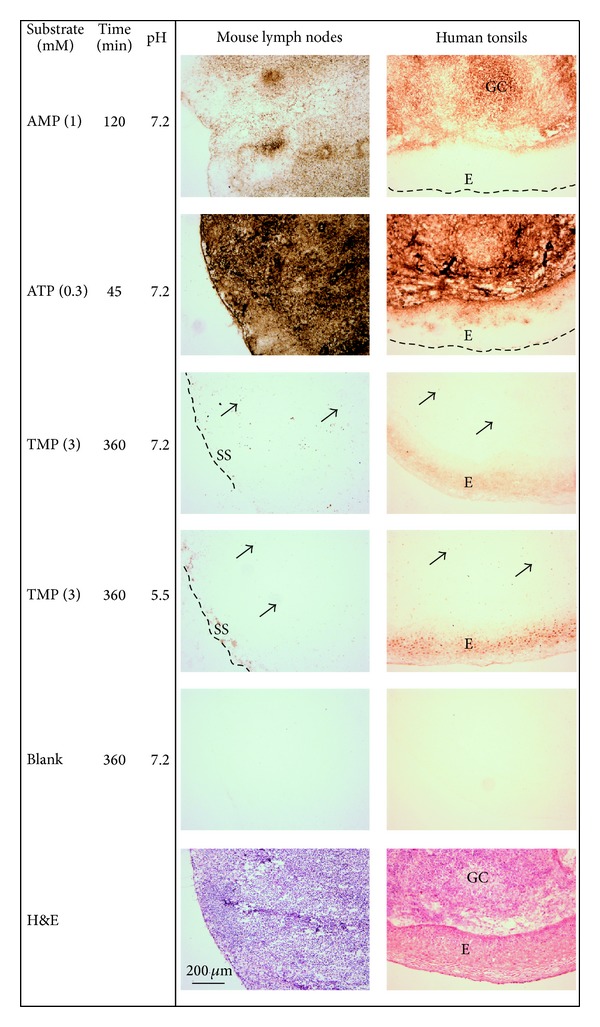
Figure 4.

Distribution of nucleotidase activities in mouse lymph nodes and human tonsils. Enzyme histochemical staining was performed by incubating tissue sections at different pH for the indicated time without (Blank) and with different nucleotide substrates. Tissue sections were also stained with hematoxylin and eosin (H&E). The capsules are indicated by dashed lines, the arrows point at some positive scattered cells, GC: germinal center, SS: subcapsular sinus, and E: epithelium. Original magnification: ×100. Scale bar: 200 μm.
