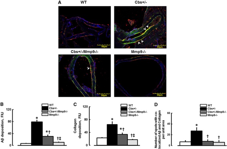
Figure 5.
Deposition of amyloid beta (Aβ) on vascular collagen in mouse brain vessels. (A) Examples of vessel images in samples obtained from wild type (WT), Cbs+/−, Cbs+/−, and matrix metalloproteinase-9 (Mmp9−/−) double knockout (Cbs+/−/Mmp9−/−), and MMP9 gene knockout (Mmp9−/−) mice. Deposition of Aβ (green) and collagen (red) and their co-localization (shown in yellow and indicated by arrowheads) was assessed by measuring fluorescence intensities of Aβ (green) and collagen (red) and number of spots with co-localized green and red colors after deconvolution of images. (B) Summary of Aβ deposition assessment. (C) Summary of collagen expression assessment. (D) Summary of Fg and Aβ co-localization assessment. *P<0.05 for all, versus WT; †, versus Cbs+/− ‡, versus Cbs+/−/Mmp9−/− n=5 for all groups.

