Figure 1.
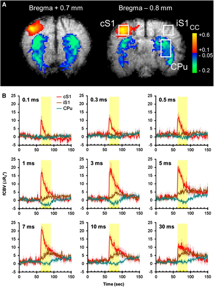
Cerebral blood volume functional magnetic resonance imaging (CBV fMRI) of right forepaw stimulation under isoflurane anesthesia. (A) Representative fMRI responses at two adjacent slices. Stimulation was 10 mA, 12 Hz, and 3 milliseconds. Stimulation-evoked CBV elevation was evident in the contralateral S1 (cS1), whereas CBV reduction was evident in the CPu of both hemispheres. (B) Group-averaged CBV fMRI time courses in the cS1, ipsilateral S1 (iS1), and ipsilateral striatum (CPu; n=11). Regions of interest are shown in A. The yellow-shaded regions indicate stimulation period. Stimuli fixed at 10 mA and 12 Hz. The intensity of the time-course data (ΔR2*) is linearly related to CBV fraction, in a unit of per second. Error bars are s.e.m. values.

