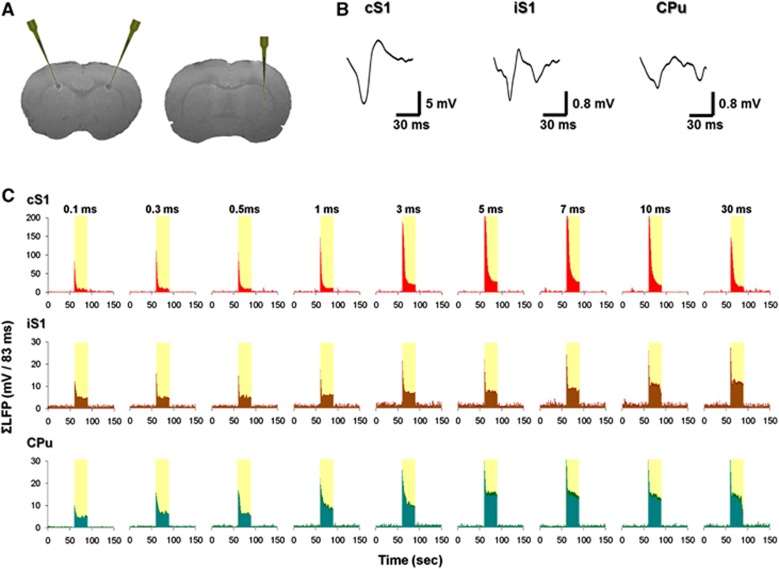
Figure 2.
Local field potential (LFP) recording of right forepaw stimulation under isoflurane anesthesia. (A) T2-weighted magnetic resonance imaging scans showing the recording site. At the end of the experiment, a 30-μA direct current was delivered to the deepest contact lead for 10 seconds for each electrode. Data were recorded at cortical layer V, ∼0.6 mm above the lesion site. (B) Typical averaged LFP waveforms in the contralateral S1 (cS1), ipsilateral S1 (iS1), and ipsilateral striatum (CPu). (C) Group-averaged ΣLFP time courses in the cS1, iS1, and CPu (n=5). Stimuli fixed at 10 mA and 12 Hz. Error bars (labeled in deeper colors) are s.e.m. values.

