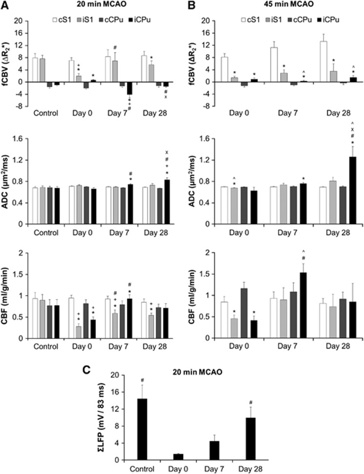
Figure 6.
CBV functional magnetic resonance imaging response, ADC, and CBF in (A) 20-minute MCAO (n=5) and (B) 45-minute MCAO group (n=5). The regions of interest are the same as shown in Figure 4B. (C) ΣLFP in 20-minute MCAO (n=4), where the recording electrode was implanted into the iCPu. P<0.05 indicates statistical significance. *different from contralateral side; +different from control; #different from day 0; Xdifferent from day 7; ^different from the matched time point in the 20-minute MCAO group. Error bars are s.e.m. values. ADC, apparent diffusion coefficient; CBF, cerebral blood flow; CBV, cerebral blood volume; cCPu, contralateral CPu; cS1, contralateral S1; fCBV, functional CBV; iCPu, ipsilateral striatum; iS1, ipsilateral S1; LFP, local field potential; MCAO, middle cerebral artery occlusion.

