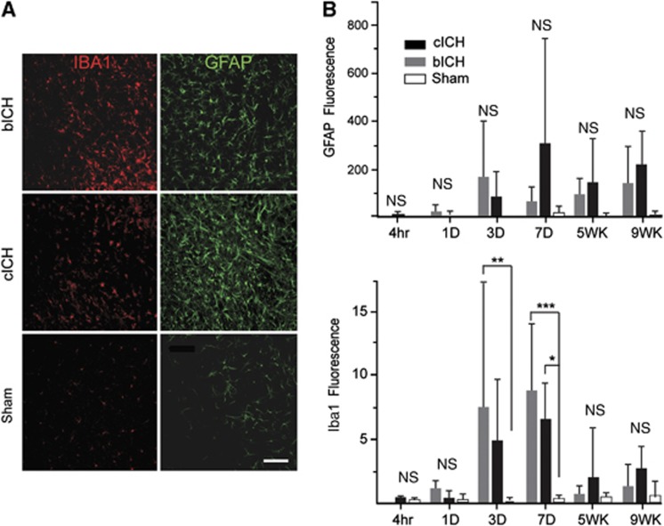
Figure 2.
Inflammatory and astrocyte responses after intracerebral hemorrhage (ICH). (A) Iba-1-positive microglia/macrophages (red) and glial fibrillary acidic protein (GFAP)-positive astrocytes (green) are visible in the perihematomal striatum in both autologous blood intracerebral hemorrhage (bICH) and collagenase intracerebral hemorrhage (cICH) at 7 days after stroke. (B) Quantification of the GFAP And Iba-1 immunoreactivity indicated that the increase in GFAP staining did not reach significance but the Iba-1 staining was greater at day 7 in bICH and cICH (n=4 per group, *P<0.05, **P<0.01, ***P<0.001). Scale bar, 100 μm. NS, nonsignificant.

