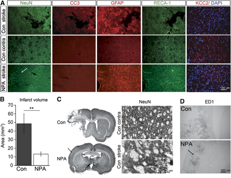
Figure 4.
Ischemic lesions after MCAO in controls (Con) and NPA-preconditioned animals. (A) Representative immunofluorescence images demonstrating expression of NeuN (green), CC3 (red), GFAP (red), RECA-1 (green), and KCC2 overlaid onto DAPI (red and blue) on the ipsilateral (stroke) and contralateral (contra) side in controls (upper and middle panel) as well as on the ipsilateral (stroke) side in NPA-preconditioned animals (lower panel). Two weeks after MCAO, tissue necrosis has occurred in controls, whereas only selective neuronal damage can be detected in NPA animals (white arrow on the lower panel NeuN staining). (B) Infarct size derived from NeuN immunohistochemistry in Con and NPA animals. Note that this analysis includes areas of cystic tissue degeneration as well as selective neuronal loss. (C) Left: overview ( × 2) of NeuN immunohistochemistry in a Con (upper panel) versus NPA (lower panel) animal. Right: higher ( × 10) magnification of the NeuN immunohistochemistry in a control animal showing normal striatal NeuN expression on the contralateral (upper panel) and loss of NeuN expression on the ipsilateral (lower panel) side. (D) ED1 immunohistochemistry demonstrating macrophage infiltration at the infarct border in controls (upper panel) and at the zones of selective neuronal death in NPA animals (lower panel). Arrows in C and D indicate areas of selective neuronal damage in NPA animals. DAPI, 4,6-diamidino-2-phenylindole 4,6-diamidino-2-phenylindole; MCAO, middle cerebral artery occlusion; NPA, 3-nitropropionic acid.

