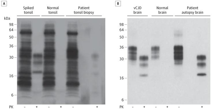
Figure 1. Immunoblot Detection of Disease-Related Prion Protein in the Patient’s Tonsil and Brain.
A, 0.5-mL aliquots of 10% weight/volume (w/v) tonsil biopsy homogenate from the patient with suspected variant Creutzfeldt-Jakob disease (vCJD) or 10% (w/v) normal human tonsil homogenate, either lacking (normal tonsil) or containing a spike of 50-nL 10% (w/v) vCJD brain homogenate (spiked tonsil), were subjected to sodium phosphotungstic acid precipitation.6 20-μL aliquots of whole samples isolated prior to centrifugation were analyzed in the absence of proteinase K (PK) digestion (−) and compared with PK-digestion products derived from the entire sodium phosphotungstic acid pellets (+). B, Aliquots of 10% (w/v) autopsy brain homogenate from the patient, a normal human control case, and a vCJD case were analyzed before (−) or after (+) PK digestion. The immunoblots were developed with anti-prion protein monoclonal antibody 3F4 using high-sensitivity chemiluminescence.

