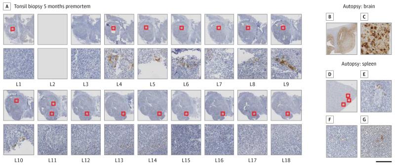
Figure 2. Immunohistochemical Detection of Abnormal Prion Protein (PrP) in the Tonsil Biopsy (A) and the Autopsy Tissues (B-G).
A, Multiple levels of the tonsil were prepared and show abnormal PrP in 2 adjacent, but separate, follicles (levels 4-6 and 9). Both follicles show moderate mechanic artifacts. Stains show an overview (B) and details (C) of the autopsy specimen of the frontal cortex illustrating substantial deposits of abnormal PrP. D, Overview of the spleen section with the 3 follicles containing PrP highlighted. High-power magnification of the same follicles to show the scarcity of PrP deposition in follicular dendritic cells (E-G). The scale bar indicates 2 mm in A; 6 mm in B and D; and 200 μm in C and E-G.

