
Figure 4.
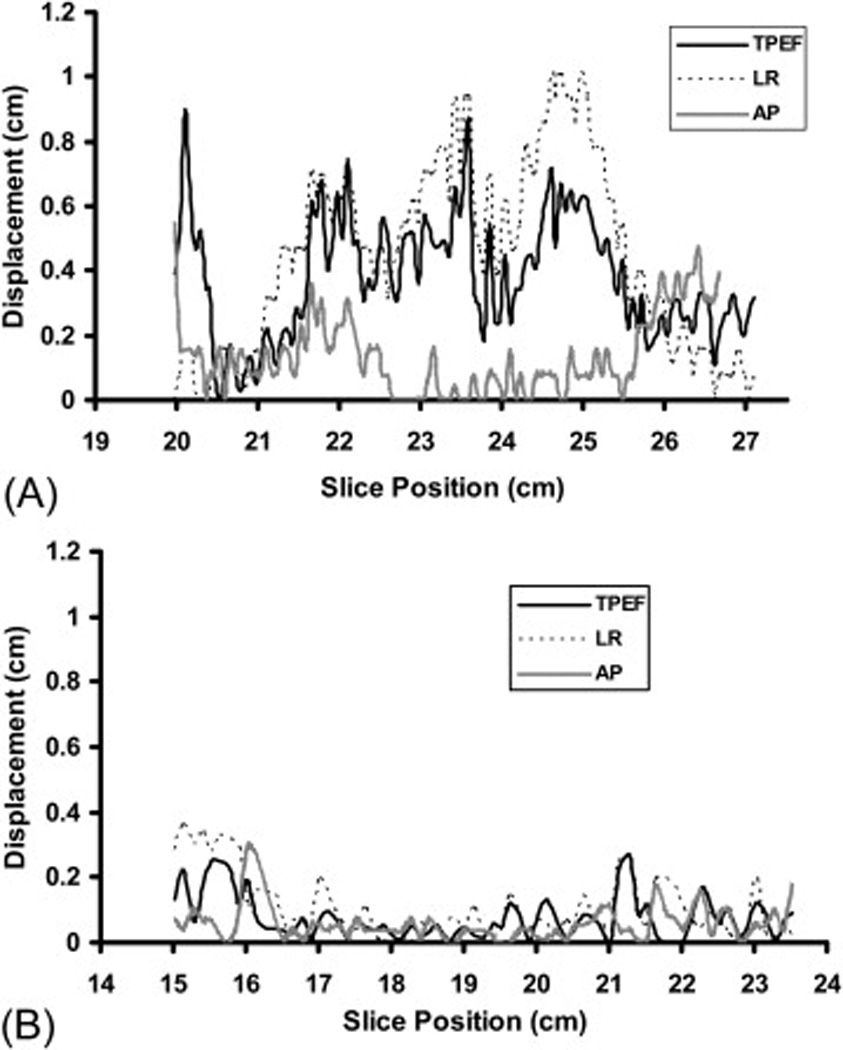
Displacement of LAD during systolic and dystolic phases of the cardiac cycle in three directions (toward the posterior edge of the treatment field [TPEF], left-right [LR], and anterposterior [AP]) on CT slices for a patient with large displacement (Panel A) and for patient with small displacement (Panel B). The × axis is the CT slice position (with smaller number indicating the superior heart and the larger number indicating inferior heart). The y axis is the displacement distance.