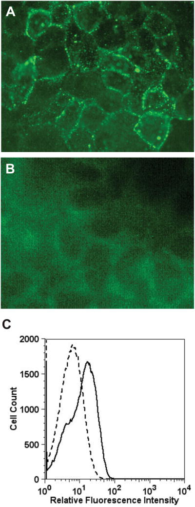
Figure 5.

Binding of GRP78 to human melanoma DM6 cells (A) Immunofluorescence microscopy of non-permeabilized DM6 cells incubated with anti-GRP78 IgG. (B) Immunofluorescence microscopy of non-permeabilized DM6 cells incubated with nonimmune human IgG. (C) Flow cytometric analysis of anti-GRP78 IgG binding to DM6 cells. The solid line represents cells incubated with autoantibodies directed against GRP78. The dashed line represents binding of isotype control.
