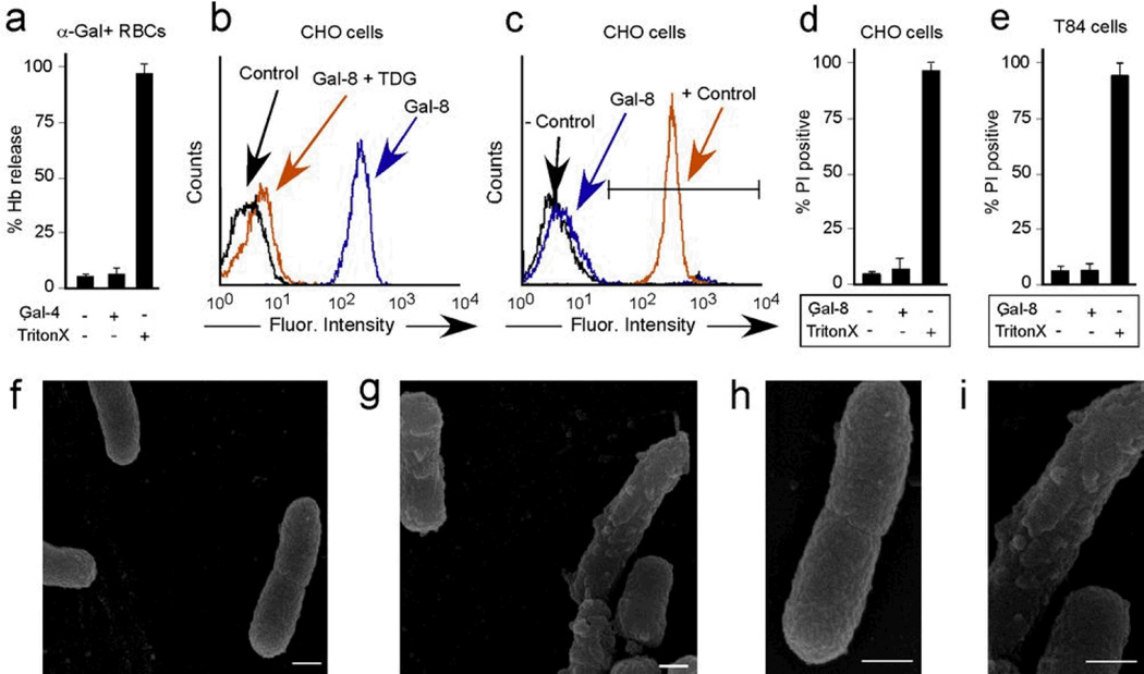
Figure 6. Galectin binding to eukaryotic cells does not alter cell viability.
(a) Quantification of hemoglobin release from rabbit erythrocytes after incubation with 5µM Gal-4 or 1% Triton X (n=2–3 in 1 representative experiment of 3). (b) Flow cytometric analysis of WT CHO cells after incubation with ~0.1µM Gal-8 with or without inclusion of 20mM TDG. (c) Flow cytometric analysis of PI positive WT CHO cells after incubation with ~0.1µM Gal-8 or 1% Triton X (+ control). (d) Quantification of percent PI+ WT CHO cells after incubation with 5µM Gal-8 or 0.1% Triton X (n=2 in 1 representative experiment of 3). (e) Quantification of percent PI+ T84 epithelial cells after incubation with 5 µM Gal-8 or 0.1% Triton X (n=3 in 1 representative experiment of 3). (f–i) Scanning electron microscopy images (15,000×) of KP O1 after incubation with PBS (f) or 5µM Gal-8 (g). Increased magnification of panels f (h) and g (i). Scale bars = 500 nm.

