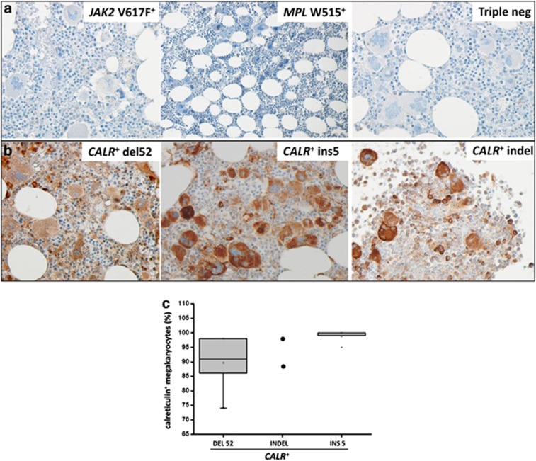
Figure 2.
Immunostaining of bone marrow biopsies with the anti-mutated CALR antibody. Representative sections from CALR-unmutated ET/PMF patients (JAK2V617F mutated, MPLW515L mutated and triple-negative mutation) and from CALR-mutated patients (CALRdel52, CALRins5 and CALRindel) are shown in panel a and panel b, respectively. Sections were stained according to the procedure described in the Materials and Methods section using a 1:1000 dilution of the anti-mutated CALR antibody. Pictures were taken with a LEICA DM LS2 microscope using a N-Plan × 40/0.65 objective. The number of immunostained megakaryocytes over total number of morphologically recognizable megakaryocytes was calculated by counting at least 100 megakaryocytes/slide from 10 patients with ET and 10 patients with PMF (n=2 for the CALR indel), and expressed as percent of total megakaryocytes (c); as there was no significant difference between the two groups, all individual data were pooled.

