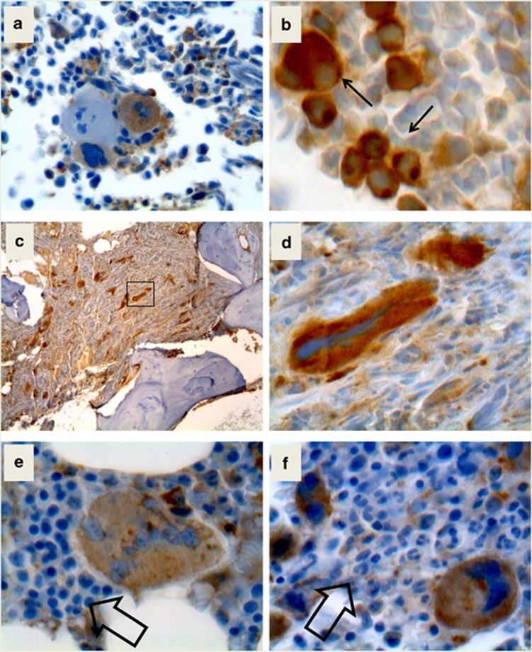
Figure 3.
Immunostaining of bone marrow biopsies with the anti-mutated CALR antibody. Panel a shows two megakaryocytes labeled by the anti-mutated calreticulin antibody together with a negative one. In panel b, abnormal, small megakaryocytes in the bone marrow of a CALRins5 PMF patient are shown. Panel c shows a low-resolution picture of an advanced fibrosis in a CALRdel52 patient and d is a high-power field from panel c (square) to show the abnormally shaped large megakaryocytes within buddles of fibers. In panels e and f, the faint label of erythroid and myeloid cells is presented at higher magnification.

