
Fig. 2.
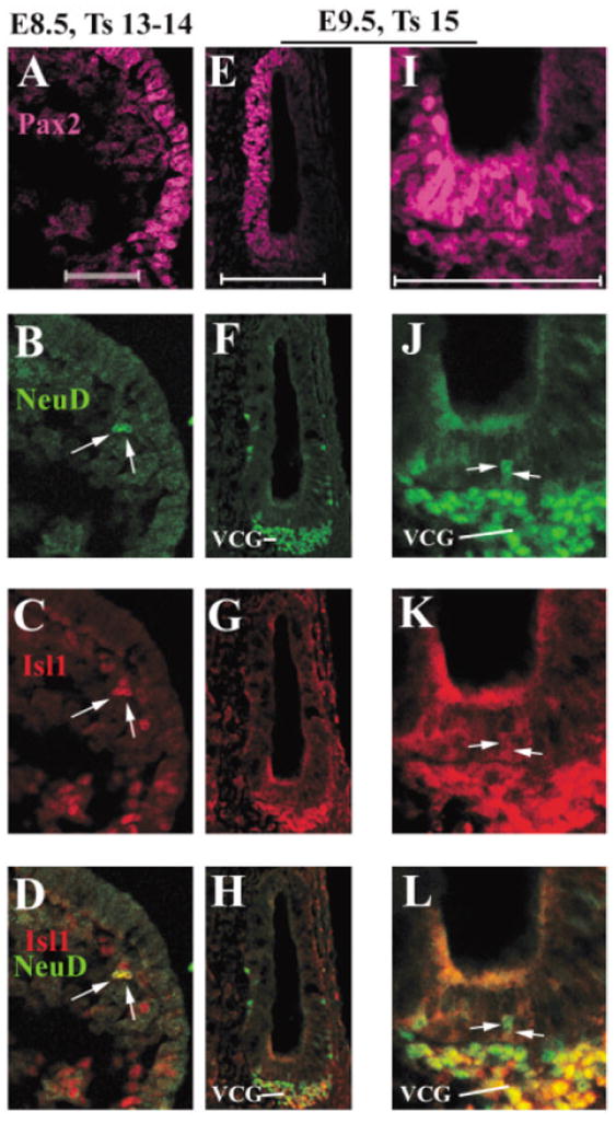
Expression of Islet1 (Isl1) in the inner ear neuronal lineage from embryonic day (E) 8.5 to E9.5. A–L: A vertical section from E8.5 (A–D) and a transverse ventral section from E9.5 (E–L) embryos were triply stained with antibodies against Pax2 (A,E,I, magenta), NeuroD (B,F,J, green), and Isl1 (C,G,K, red). D,H,L: Overlays of NeuroD and Isl1 staining. I,J,K,L: Higher magnified images of E,F,G,H, respectively. For E8.5 vertical section, dorsal is at the top, and for E9.5 transverse section, anterior is at the top. Medial is at the left for both the sections. VCG, vestibular–cochlear ganglion neurons. Scale bars = 50 μm in A (applies to A–D), E (applies to E–H), I (applies to I–L).