
Fig. 3.
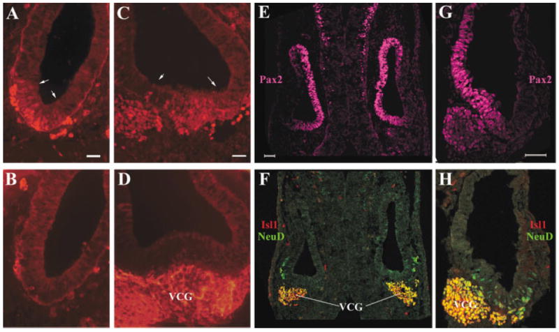
Expression of Islet1 (Isl1) in the inner ear neuronal lineage at embryonic day (E) 10.5. A–D: Adjacent vertical (A,B) or transverse (C,D) E10.5 sections were stained for Isl1 (A,C) or Tuj-1 (C,D). Arrows in A and C delimit the expression domain of Isl1 in the otic epithelium at this stage. E–H: Transverse (E,F) and vertical (G,H) sections were triply labeled for Pax2 (E,G, magenta), Isl1 (F,H, red), and NeuroD (F,H, green). F,H: The staining for Isl1 and NeuroD was overlaid. VCG, vestibular–cochlear ganglion neurons. Scale bars = 50 μm in A (applies to A,B), C (applies to C,D), E (applies to E,F), G (applies to G,H).