
Fig. 6.
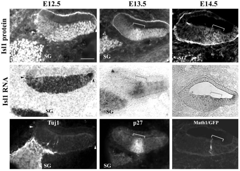
Islet1 (Isl1) expression in the cochlea. A–I: Adjacent cochlear sections from embryonic day (E) 12.5 (A–C), E13.5 (D–F), and E14.5 (G–I) stained for Isl1 antibody (A,D,G), Isl1 mRNA (B,E,H), neuronal β-tubulin antibody (C, Tuj1), p27Kip1 antibody (F), or with Math1/GFP signal (I). Arrowheads in A–C mark the boundaries of the ventral cochlear epithelium expressing Isl1 at E12.5. Brackets (D–I) indicate the primordial organ of Corti before terminal differentiation. SG, spiral ganglion neurons. Medial is at the left of the images. GFP, green fluorescent protein. Scale bar = 50 μm in A (applies to A–I).