
Fig. 7.
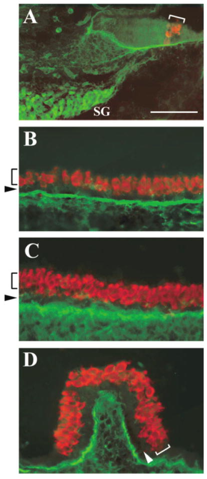
Differential expression of Islet1 (Isl1) in the sensory and neuronal cells of the inner ear. A–D: Cross-sections of the cochlea (A), saccule (B), utricle (C), and crista (D) at embryonic day 16.5 double stained for Isl1 (A, green) and Math1/GFP (A, red) antibodies, or Isl1 (B–D, green) and myosin VIIa (B–D, red) antibodies. The brackets indicate the organ of Corti in the cochlea (A), or the hair cell layer in the vestibular sensory organs (B–D). The arrows indicate the supporting cell layer in the vestibular sensory organs (B–D). Isl1 is absent in the sensory organs but persists in SG neurons. SG, spiral ganglion neurons; GFP, green fluorescent protein. Scale bar = 50 μm in A (applies to A–D).