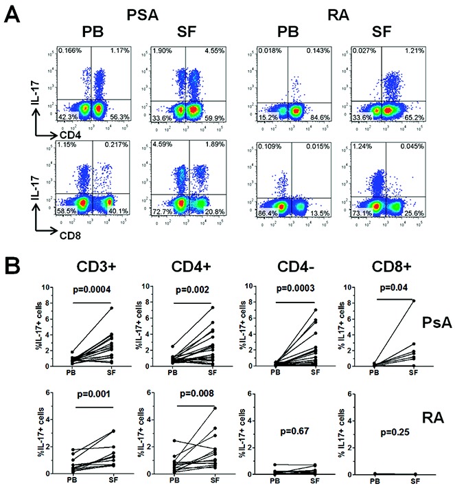
Figure 1.

Frequency of interleukin-17 (IL-17)–expressing cells in CD3+, CD3+CD4+, CD3+CD4−, and CD3+CD8+ T cell populations in paired peripheral blood (PB) and synovial fluid (SF) samples from patients with psoriatic arthritis (PsA) or rheumatoid arthritis (RA). Mononuclear cells from paired PB and SF samples from patients with PsA (n = 21) and patients with RA (n = 14) were isolated and stimulated as described in Patients and Methods. A, Percentage of IL-17–expressing cells within CD4+ and CD4− T cells (top panels) and within CD8+ and CD8− T cells (bottom panels), as determined by flow cytometry in PB and SF from a representative patient with PsA and a representative patient with RA. B, Percentage of IL-17+ cells in total CD3+, CD4+, CD4−, or CD8+ T cells in paired PB and SF samples from patients with PsA and patients with RA. For the CD8+ subset, the percentage of IL-17+ cells was determined in paired samples from 8 PsA patients and 3 RA patients. Data were analyzed using Wilcoxon's matched pairs signed rank test. Color figure can be viewed in the online issue, which is available at http://onlinelibrary.wiley.com/doi/10.1002/art.38376/abstract.
